Osteology 101- Part 4/5: Paleopathology
- Aug 24, 2019
- 4 min read
Sometimes, we bonk our head a little too hard. We'll maybe joke, “Ouch, that’ll leave a mark”. But the osteological question is, can head bonks, falls, and drawn-out sicknesses actually show up on your skeleton? These types of inquiries fall into the study of paleopathology. Paleopathology is the study of ancient diseases, which can include infection, trauma, autoimmune disorders, and dental pathology, to name a few. Though we could spend ages going through every possible pathology that can be visible on the skeleton, today’s intention is not to try to put you to sleep, therefore, we will cover only a few of the possibilities.
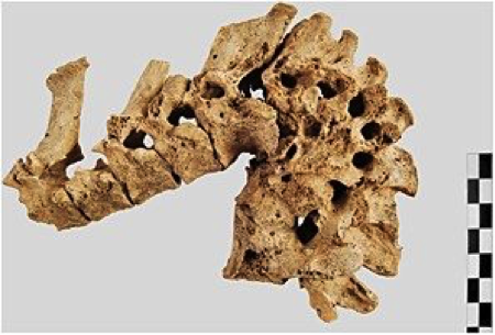
Infectious diseases are when the biological body is invaded with disease-causing agents. Eventually the organism will react to the infection, which is when some infections begin to be skeletally observable. In today’s video, you saw some examples of tuberculosis (TB) and syphilis. TB is caused by Mycobacterium tuberculosis bacteria and is an airborne disease. It typically begins by attacking the lungs, hence the coughing and sneezing that will follow. Once the infection has ravaged the body, the skeleton will show signs of vertebral fusion and collapse, joint ankylosis (fusing) and destruction, to the point of rib periostitis. It ain’t pretty, I’ll tell you that..


On the other hand, treponemal diseases: yaws, pinta, venereal and endemic syphilis; have many forms and varying skeletal indicators. Though for the sake of general understanding of paleopathology, it is interesting to consider syphilis, as an example. It has been suggested, that syphilis was not spread through sexual contact in the past but skin-to-skin, like yaws. When sanitation conditions improved, the disease mutated to become the sexually transmitted disease we know it as today. These factors are crucial to know of diseases evolving over time, as humans do. Thus, the study of paleopathology can improve the understanding of modern diseases by acknowledging their development over time. There is value to history!

Syphilitic Skull
Osteological trauma refers to a wound in the skeleton that is caused by external force. This can be as simple as a bone fracture or break, to a more extreme application of trauma from a projectile, object, or accident producing projectile, blunt-, or sharp-force trauma. There are several types of bone fractures, which will heal differently depending on their type, location, extremity, age of individual, and time before death (assuming they did not play a role in the cause of death).

Different types of fractures
The other traumas relate to circumstances such as hits and weapons. These injuries alter the bone, and depending on the timing of the trauma, this can show the osteologist the possible circumstances the individual may have experienced whilst alive.

Gunshot entry and exit wound
Autoimmune disorders can include many conditions where the immune system attacks the body (e.g. psoriasis, arthritis, Graves’ disease, Celiac disease), though not all will be visible on the skeleton. Arthritis is one of the most obvious to the osteologist and can be result of several autoimmune diseases. We will only discuss Osteoarthritis (OA) today.

Process of osteoarthritis
OA is a degenerative disease. It occurs when the protective cartilage that cushions the ends of the bones wears down over time. This can be a cause of old age, joint injuries or repeated stress on the joints, genetics, metabolic diseases, and bone deformities. The joints and the adjacent bone will form bony lipping and spur formation around the joint edges and can also develop eburnation (i.e. polishing) on the surfaces. Though OA is interesting to observe, activity-related OA can provide information about the life hood of the individual. This could entail their job, activities, and/or roles in society (i.e. hunter, sports, basket weaver, etc).

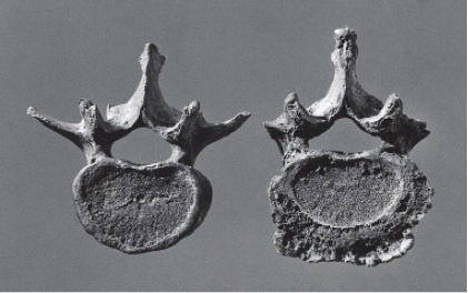

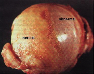
OA in the vertebra Eburnation or polishing of the femoral head
Lastly, for today, dental pathologies. Dental pathologies include things such as caries (i.e. cavities), enamel hypoplasia (i.e. developmental defects), periodontal diseases, antemortem tooth loss (AMTL). Enamel hypoplasia can affect both the baby and adult teeth and is a defect more commonly recognised to occur from vitamin deficiencies, but also developmental instabilities. It results in the enamel being deficient in amount. EH indicators can appear as lines, pits, or grooves in the crown of the tooth.

[Linear] Enamel Hypoplasia in the lower adult teeth
Cavities are decayed areas of the teeth that develop into openings, one of the most commonly known dental disease to all of us. Furthermore, periodontal diseases are infections around the teeth that begins with gingivitis (plaque build-up or ‘calculus’) and can lead to periodontitis, where one can prematurely lose teeth (AMTL).
Of course, we could have discussed scoliosis, rickets, gout, cancer, trepanation, etc. However, the study of Paleopathology in Osteology is a meticulous field, that only grows as we better understand the origins and development of diseases.


Top Left: Scoliosis vs, normal spine.
Top Right: Radiograph of scoliosis.
Left: Normal lower body vs, ricket lower body.
Naturally, seeing the diseases affect the bone is much more effective than reading about it. Therefore, today I have provided you with several photos that can give you a better idea of what bones show us. Now go into the world, and try not to think about what your skeleton might have, too much...
References


toto slot, toto slot, toto slot, toto slot, toto slot, raja168, raja168, raja168, raja168, raja168, toto slot, toto slot, toto slot, toto slot, toto slot, toto slot, toto slot, toto slot, toto slot, raja168, raja168, toto slot, toto slot, toto slot, toto slot, raja168,
slot gacor, slot gacor, slot gacor, slot gacor, slot gacor, slot gacor, slot gacor, raja168, raja168, raja168, raja168, raja168, raja168, slot gacor, slot gacor, slot gacor, slot gacor, slot gacor, slot gacor, slot gacor, raja168, raja168, raja168, slot gacor, slot gacor, slot gacor, slot gacor, slot gacor, slot gacor, slot gacor, slot gacor, slot gacor, slot gacor, slot gacor, raja168, slot gacor, raja168, raja168, slot gacor, slot gacor, slot gacor, slot gacor, slot gacor, raja168, raja168, slot gacor, raja168, slot gacor, slot gacor, slot gacor, slot gacor, slot gacor, raja168,